PRP
What is PRP Therapy?
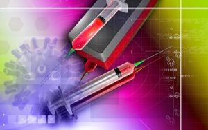
PRP is a new natural therapy that has been increasingly used in various disorders especially for the last 5 years. Although it has originated as a cosmetic and anti-aging therapy, it is recently used in the treatment of musculoskeletal system disorders. PRP stands for “Platelet Rich Plasma”. The medication is prepared from the patient’s own blood.
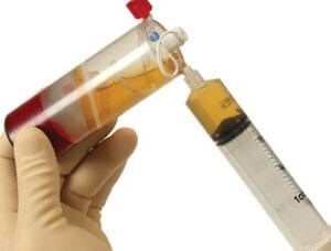
How to Obtain PRP
Approximately 10-20 cc of blood is drawn from the patient, based on the application. This blood is processed in specifically designed systems. The blood cells of the patient is centrifuged to achieve a platelet rich plasma fluid on the upper layer.
The Effect of PRP in The Body
Platelet is mainly the cell that provides coagulation and includes growth factor and some natural protectors. Platelet rich plasma operates as a natural medication. It speeds up the healing process of injured structures such as tendon, cartilage.
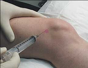
How Administered
It is injected into the treatment site 2-3 times usually with one month intervals with doses of 1-3 ml.
How to provide treatment for the patient with knee arthrosis Read more
PRP Application Areas
Knee osteoarthritis (Gonarthrosis): It is usually administered 3 times with monthly intervals.
Watch PRP Application
How to treat osteoarthritis of the hip joint? Read more
In the cases of osteoarthritis of the hip, shoulder and other joints: It is usually administered 3 times
Watch how to administer prp shoulder injection
Chronic tendinitis (tennis elbow, golfer’s elbow): It is usually administered 2 times.
Tendon injuries (Achilles tendinitis, adductor tendinitis, sports injuries): It is administered 1-3 times.
Rotator cuff tears: It is administered 1-2 times.
Are There Any Side Effects of PRP?
PRP method has no side effects. It may only cause temporary pain and swelling in the treatment site. This will go away in a couple of days and will not cause any permanent damage.
PRP Treatment in Ankle Cartilage Injury
Ankle sprain is one of the most common musculoskeletal system issues. This can occur both in athletes and ordinary people. One can experience ankle sprain both due to constraining in sports activities and also descending a ladder. Football matches are among the most common reasons for ankle cartilage problems in men. The fields with high friction is especially a great danger for under-exercised players. While walking on high heels is among the most common reasons for women.
Ankle is twisted inwards, in almost all cases, and this causes various levels of injuries on the lateral ligaments of the ankles. In mild injuries, the lateral ligaments are stretched. In severe injuries, these ligaments are torn. Patients may experience swelling and bruises on especially outer side of the ankle. The patient experiences pain when stepping on and has limited capacity of ankle movements. Usually putting ice on, resting, bandage and pain killers would provide sufficient efficacy in mild injuries. However, for severe injuries, the ankle may need to be stabilized with a brace or basic casts. Following the acute phase, physical therapy should be provided to accelerate recovery.
Patients with ankle sprains may experience a serious issue: A regional damage might occur on the articular cartilage of the ankle due to sprains, and this condition is called Osteochondritis. In “Talar Dome”, there is an articular cartilage injury and underlying bony involvement in the form of edema. In some cases, the injured part of the cartilage crumbles and bone is exposed in that area. Due to the deficiency of cartilage, corrosion starts in bones over the time and ankle arthritis, which is normally rare, occurs. This leads to permanent pain. In rare cases, a cartilage piece may fall into the joint spacing following ankle sprain. It requires arthroscopic removal procedure. If the patient experience permanent pain after the acute phase of ankle sprain, especially when stepping on and walking for an extended period of time, Osteochondritis should be taken into consideration. Osteochondritis in the ankle can only be diagnosed with magnetic resonance imaging (MRI). MRI examination also shows the degree of the injury.
Physical therapy can contribute to recovery of Osteochondritis by reducing the intra articular edema. Na hyalurinate injections administered intra-articularly into the ankle joint, increase the viscosity of the joints and prevents a certain level of damage to the joint.
Recently PRP therapy, prepared from the patient’s blood, is administered in cases of Osteochondritis. PRP stands for platelet rich plasma. PRP , concentration of platelets that are extracted from autologous blood, supports the injured cartilage to prevent crumbles in especially early phases of Osteochondritis, preventing ankle osteoarthritis in the long term. PRP is administered into the ankle joint at monthly intervals, 3 times in total, under USG. PRP may also be administered into the injured ligaments. PRP accelerates the recovery of the ligaments injured and strengthens the ligaments.
In all cases, special exercises for ankle should be done to support both PRP or other treatment methods. This can prevent a second ankle sprain, which is another great danger for these patients. If patient does not do preventive exercises, 30-70% of the cases the patient experiences a second ankle sprain. This may get so frequent to be defined as “chronic ankle sprain”. In this condition, the ankle loses its stability. In late phases, it may require surgical intervention.
Everyone may have an ankle sprain. The important thing is to take preventative cautions to avoid relapse. Osteochondritis should be taken into consideration in case of long term pain following the ankle sprain. Early diagnosis and the right treatment method can minimize the harm of Osteochondritis.
Plantar fasciitis, meniscal tear, osteochondritis, chronic bursts, ankle strain are among other areas of usage.